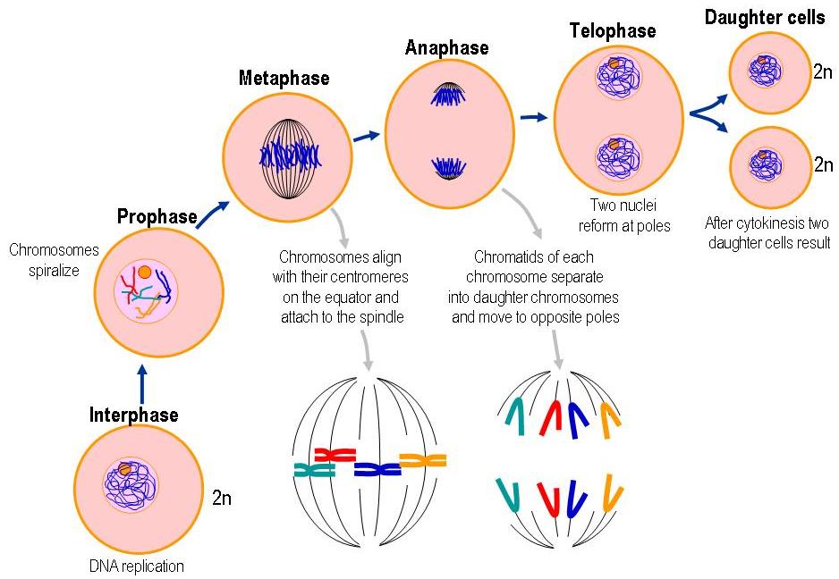

They are referred to as daughter chromosomes. Once the paired sister chromatids separate from one another, each is considered a "full" chromosome.The paired centromeres in each distinct chromosome begin to move apart..During anaphase, the following key changes occur: At the end of anaphase, each pole contains a complete compilation of chromosomes. Spindle fibers not connected to chromatids lengthen and elongate the cell. In anaphase, the paired chromosomes ( sister chromatids) separate and begin moving to opposite ends (poles) of the cell. The chromosomes begin to migrate toward the cell center.The kinetochore fibers "interact" with the spindle polar fibers connecting the kinetochores to the polar fibers.Kinetochores, which are specialized regions in the centromeres of chromosomes, attach to a type of microtubule called kinetochore fibers.Polar fibers, which are microtubules that make up the spindle fibers, reach from each cell pole to the cell's equator.The two pairs of centrioles (formed from the replication of one pair in Interphase) move away from one another toward opposite ends of the cell due to the lengthening of the microtubules that form between them.The mitotic spindle, composed of microtubules and proteins, forms in the cytoplasm.

Chromatin fibers become coiled into chromosomes, with each chromosome having two chromatids joined at a centromere.During prophase, a number of important changes occur: Prophase (versus interphase) is the first true step of the mitotic process. The nuclear envelope breaks down and spindles form at opposite poles of the cell. Then, in the second part of anaphase - sometimes called anaphase B - the astral microtubules that are anchored to the cell membrane pull the poles further apart and the interpolar microtubules slide past each other, exerting additional pull on the chromosomes (Figure 2).In prophase, the chromatin condenses into discrete chromosomes. More specifically, in the first part of anaphase - sometimes called anaphase A - the kinetochore microtubules shorten and draw the chromosomes toward the spindle poles. Meanwhile, changes in microtubule length provide the mechanism for chromosome movement. Upon separation, every chromatid becomes an independent chromosome. Enzymatic breakdown of cohesin - which linked the sister chromatids together during prophase - causes this separation to occur. Metaphase leads to anaphase, during which each chromosome's sister chromatids separate and move to opposite poles of the cell. Kinetochore microtubules attach the chromosomes to the spindle pole interpolar microtubules extend from the spindle pole across the equator, almost to the opposite spindle pole and astral microtubules extend from the spindle pole to the cell membrane. In addition, the spindle is now complete, and three groups of spindle microtubules are apparent. At this point, the tension within the cell becomes balanced, and the chromosomes no longer move back and forth. Every chromosome has at least two microtubules extending from its kinetochore - with at least one microtubule connected to each pole. This process ensures that each daughter cell will contain one exact copy of the parent cell DNA.Īs prometaphase ends and metaphase begins, the chromosomes align along the cell equator. As they move, they pull the one copy of each chromosome with them to opposite poles of the cell. The spindle tubules then shorten and move toward the poles of the cell. As mitosis progresses, the microtubules attach to the chromosomes, which have already duplicated their DNA and aligned across the center of the cell. These tubules, collectively known as the spindle, extend from structures called centrosomes - with one centrosome located at each of the opposite ends, or poles, of a cell. Early microscopists were the first to observe these structures, and they also noted the appearance of a specialized network of microtubules during mitosis. The word "mitosis" means "threads," and it refers to the threadlike appearance of chromosomes as the cell prepares to divide. Mitosis is the process in which a eukaryotic cell nucleus splits in two, followed by division of the parent cell into two daughter cells.


 0 kommentar(er)
